Publicaciones

Perfil ORCID

Artículos y tesis

AñoFiguraTítulo · autores · fuenteIdentificador
2026
Parietal thickness and Homo antecessor: a description of three new fragments from Gran Dolina, Atapuerca, SpainGallareto-Sande, R., Bruner, E., Saladié, P., Martinón-Torres, M.Journal of Human Evolution, 218, 103870
Resumen

Homo antecessor, from the Early Pleistocene site of Gran Dolina (Atapuerca, Spain), occupies a basal phylogenetic position within the clade that includes Neanderthals and modern humans, yet its cranial morphology remains poorly characterized due to the fragmentary nature of the preserved remains. Here we describe three previously unstudied parietal fragments (ATD6-115, ATD6-128, ATD6-401) from the TD6 level, representing a minimum of two adult individuals, together with the juvenile specimen ATD6-100/168. Using micro-computed tomography, we quantified total cranial vault thickness (CVT) and the relative contributions of the external table, diploic space, and inner table at 20 standardized points per specimen. Mean total CVT in the adult fragments ranges from 6.07 mm to 7.03 mm, whereas the juvenile specimen is thinner (4.65 mm). In all specimens the diploic space constitutes the dominant layer (40–50% of CVT). Comparative analysis with published data places the adult H. antecessor parietal thickness within the range of H. neanderthalensis and modern humans, and below the robust condition of our comparative Early Pleistocene Homo sample. The spatial distribution of thickness follows a decreasing gradient from the posterior sagittal region inferiorly, an arrangement more consistent with the variable pattern reported for modern humans than with the more uniform distribution described for Neanderthals. These features reveal a mosaic morphology combining a derived, gracile vault with primitive traits, consistent with its proposed phylogenetic position.

2026
Vascular microforamina: a preliminary quantitative study on prevalence and cranial correlates in adult humansGallareto-Sande, R., Mena-Bock, A. L., Bruner, E.The Anatomical Record
Resumen

Microforamina are small vascular channels connecting the cranial diploe to the endocranial space, hypothesized to play a role in thermoregulation and neuroimmune functions, yet their quantitative anatomy is poorly documented in humans and, in general, in vertebrates. This preliminary study provides the first analysis of their number, size, and correlation with skull thickness in 10 modern human crania, using high‐resolution industrial tomography (0.25 mm voxel size). Results show substantial interindividual variability, with an inverse relationship between channel size and frequency. Although the smallest channels were most numerous, the larger ones contributed to a comparable total luminal area. Medium‐sized channels show statistically significant correlations with cranial thickness: negative correlations with inner table thickness and positive correlation with diploic space proportion, specifically in the parietal and occipital bones. These preliminary findings establish a quantitative baseline, indicating that microforamina prevalence is more influenced by the thickness of the vault layers than with general cranial size.

2026
Morphological variations and cortical atrophy of the precuneus in normal aging and Alzheimer's diseaseBruner, E., Gallareto-Sande, R., López-González, F. J., Zhang, L., Sánchez Martín, C., Grothe, M. J., Sánchez-Juan, P.Journal of Anatomy, 248(1), 62–70★ Journal of Anatomy Runner-Up Best Paper Prize 2025 · Ver diploma
Resumen

The precuneus is a central hub of the human brain, and its morphology displays a noticeable individual variability. It is larger in humans than in other primates, and it is affected by atrophy and hypometabolism in the early stages of Alzheimer's disease (AD). In this study, we compare its general morphology in normal aging subjects and AD patients, considering its geometry through shape analysis, and the atrophy at its boundary by quantifying the intersulcal spacing. Results suggest that the AD sample displays a reduction of the inferior regions of the precuneus, namely those that fade into the posterior cingulate and retrosplenial cortex. During aging, sulcal spacing is pronounced at the superior and posterior regions, although in the AD sample the latter change is more marked. However, these differences are associated with a remarkable individual variation and a consistent overlap between group ranges. This study stresses further the differences between the dorsal and ventral regions of the precuneus. Beyond functional factors, the dorsal areas represent a region of scarce spatial constraints, whereas the ventral regions are embedded in a complex topological environment. This structural difference must be considered when investigating cortical morphology in both normal and pathological conditions.

2025
Growth and development of the precuneus in humans from birth to early adulthoodBruner, E., Gallareto-Sande, R.Cerebral Cortex, 35(12), bhaf305
Resumen

The precuneus is a main node of the brain organization, and it is crucial to autonoetic consciousness, body awareness, imagination, and the construction of the self. In humans, it displays a remarkable individual variation, which functional consequences are not yet known. Comparative analyses suggest that the expansion of these areas may have had an evolutionary relevance in Homo sapiens. In this study, we analyzed the MRI scans of 220 individuals ranging from birth to early adulthood, to investigate the morphological changes of the precuneus along ontogeny. Size changes are only detected between birth and 2–3 years of age. In terms of shape, there is a minor increase in dorsal expansion in the same period, although the mean effect is moderate, and general trends are obscured by the pronounced variability. The remarkable individual differences in precuneus morphology are already established at birth. The dorsal extension is a major determinant of such variability in all age groups, although the analysis reveals multiple factors involved. Importantly, the dorsal and ventral regions of the precuneus are largely independent. Longitudinal studies will be necessary to test the details of its morphological changes through ontogeny, as well as surveys investigating its development in prenatal stages.

2023TFM
Actividad electrodérmica y variación en la atención fásica durante la exploración háptica de bifaces y cantos tallados del Paleolítico inferiorGallareto-Sande, R.Máster en Evolución Humana, Universidad de Burgos
Resumen

La arqueología cognitiva intenta acceder a los procesos mentales de aquellos individuos extintos a través de su cultura material. Las herramientas líticas representan la mayor parte de esta cultura material para el Paleolítico inferior. La complejidad humana está fuertemente ligada a su inteligencia y esta, a su vez, al sistema atencional. Este trabajo se basa en el análisis de la atención fásica en 46 individuos adultos, durante la manipulación de 21 herramientas líticas diferentes, hasta alcanzar una posición confortable. Para esto se realizó el conteo de picos de respuesta electrodermal durante el tiempo total de la exploración háptica para cada sesión y se compararon resultados por tipología lítica. Se observó una respuesta diferencial para las herramientas de tipo achelense cuya causa estaría vinculada a la mayor complejidad técnica de esta tipología.

2022TFM
Aproximación al concepto de autocontrol en humanos. Una mirada evolutivaGallareto-Sande, R.Máster en Antropología Física y Forense, Universidad de Granada
Resumen

A través de una revisión sistemática se realiza una aproximación al concepto de autocontrol en humanos desde una mirada evolutiva, presentando los procesos cognitivo-afectivos involucrados y la importancia, e influencia, de estos en la sociabilidad. La interrelación de mecanismos, habilidades cognitivas y procesos emocionales hace muy difícil el consenso científico en cuanto a terminología y taxonomía. Aun así no hay duda de que el control consciente de los impulsos —en pos de una recompensa futura mayor— se presenta como fundamental en la construcción de vínculos que habilitan el trabajo cooperativo y la formación de grupos sociales.

Presentaciones y pósters

AñoFiguraTítulo · autoresEvento
2026Pecha Kucha
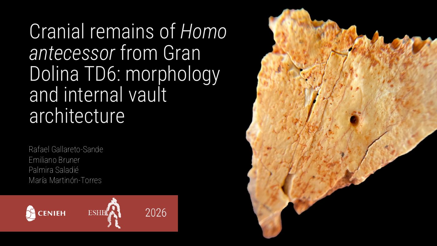
Cranial remains of Homo antecessor from Gran Dolina TD6: morphology and internal vault architectureGallareto-Sande, R., Bruner, E., Saladié, P., Martinón-Torres, M.
Resumen

Homo antecessor is one of the earliest hominin taxa documented in Western Europe, represented almost exclusively by the fossil assemblage recovered from the Aurora Stratum (TD6.2) of Gran Dolina, Sierra de Atapuerca (Burgos, Spain), dated to approximately 850 ka. Despite the taxonomic and evolutionary significance of this species, its cranial morphology remains incompletely characterized. The TD6 cranial assemblage is fragmentary, and several specimens have yet to receive formal morphological description. Here we present a comprehensive overview of the available cranial material, with the aim of advancing the anatomical characterization of H. antecessor and identifying morphological patterns relevant to its phylogenetic assessment.

The core sample comprises four parietal fragments –ATD6-115, ATD6-128, ATD6-401, and ATD6-100/168– representing a minimum of two adult and one subadult individual. ATD6-100/168 was first described and analyzed by Bruner et al. (2017) and subsequently reanalyzed alongside the three newly described specimens using a standardized protocol to ensure comparability. The morphological description and CVT analysis of all four parietals are presented in a companion study currently under revision. ATD6-60, a pterion fragment, is introduced as supplementary material. Given the structural fragility of the pterion region, its preservation as a fossil specimen is itself noteworthy, and it provides access to a portion of the cranial vault largely undocumented in H. antecessor. Morphological description and morphometric comparison with a modern human reference sample complement the parietal analysis and broaden the anatomical scope of the cranial characterization of the species.

Morphological description is based on direct physical observation and high-resolution micro-computed tomography (µCT). For the parietal sample, µCT data were used to quantify CVT and its three internal components –external table, diploic space, and inner table. Results are discussed in the context of known CVT variation across the genus Homo and are consistent with the proposed phylogenetic position of H. antecessor as a stem member of the Neanderthal–modern human clade, while underscoring the need for caution given the limited number of available cranial remains.

Taken together, this material constitutes the most complete cranial sample of H. antecessor subjected to systematic morphological analysis to date. The ongoing study of additional fragments, combined with material expected from future excavation campaigns at Gran Dolina, will continue to refine our understanding of the cranial biology of this key Early Pleistocene hominin.

European Society for the study of Human Evolution (ESHE)
Burgos, España
2025Póster
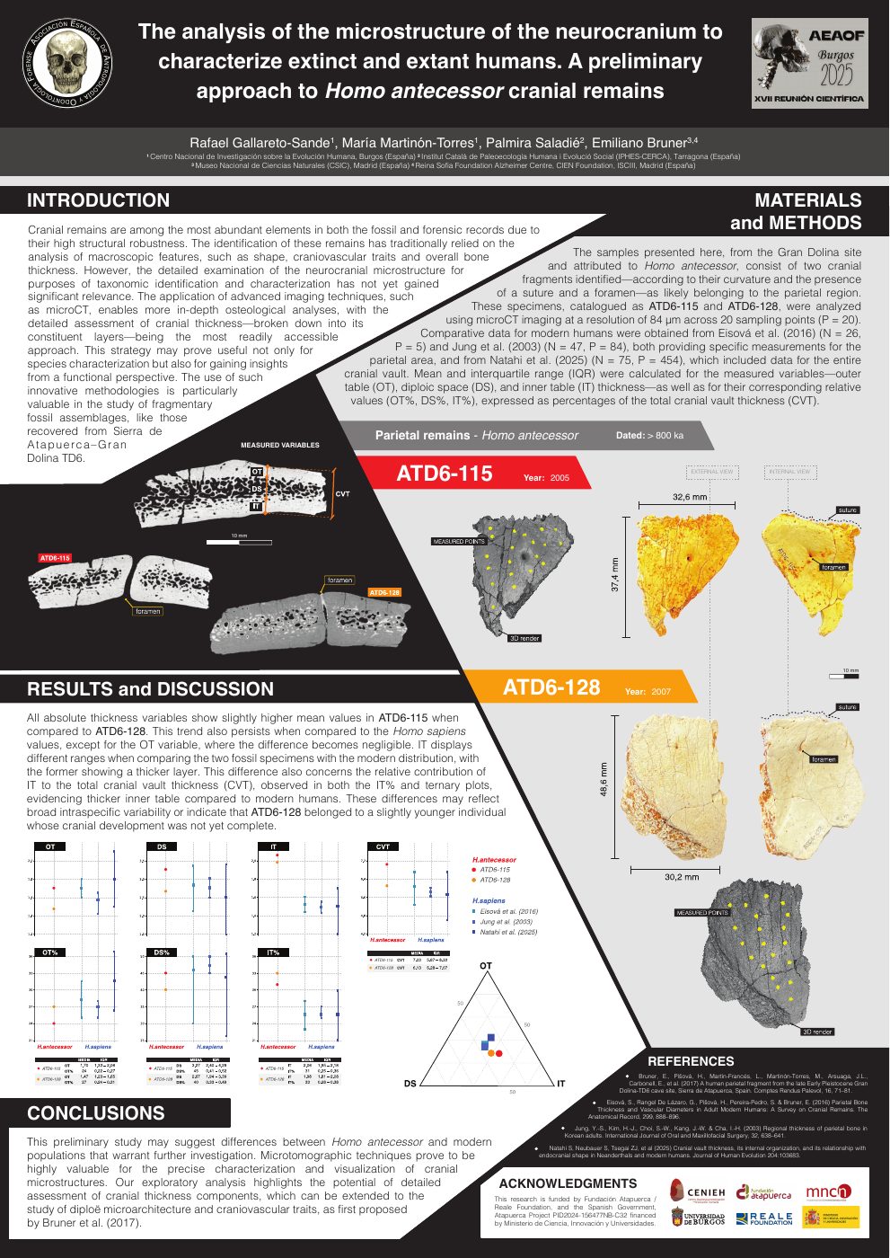
The analysis of the microstructure of the neurocranium to characterize extinct and extant humans. A preliminary approach to a H. antecessor cranial remainGallareto-Sande, R., Martinón-Torres, M., Saladié, P., Bruner, E.
Resumen

Introduction: The identification of cranial remains in both paleontological and forensic contexts has traditionally relied on macroscopic anatomical features. However, the potential of the diploë in the neurocranium to characterize both modern and extinct populations remains underexplored. The application of advanced imaging techniques, such as microCT, enables precise analysis of bone histology, paving the way for new methods of comparing cranial vault bones. This approach may prove useful not only for species characterization but also for gaining insights from a functional perspective. Such innovative methodologies are particularly valuable in the study of fragmentary fossil assemblages, like those recovered from Atapuerca–Gran Dolina TD6.

Objectives: This study presents a description of a new cranial remain of Homo antecessor through both external and internal examinations using microtomographic techniques. As a preliminary approach, the variation in neurocranial thickness—specifically the inner table, diploë, outer table, and total thickness—is compared to a reference sample compiled from the literature.

Materials and Methods: Cranial thickness data for modern humans were obtained from Eisová et al. (2016) (N=26), Beaudet et al. (2018) (N=10) and Natahi (2025) (N=85). The same type of analysis was performed on a microCT scan (0.09 mm) of a fossil cranial fragment of Homo antecessor.

Results and Discussion: Our preliminary study reveals some differences between H. antecessor and modern populations that deserve further exploration. Microtomographic techniques are useful for the precise characterization and visualization of microscopic features. Our exploratory study emphasizes the potential of detailed analysis of the cranial structure—such as diploë microarchitecture and craniovascular traits (Bruner et al., 2017)—as a valuable tool for species identification in both paleontological and forensic contexts.

XVII Reunión Científica de la AEAOF
Burgos, España
2025Póster
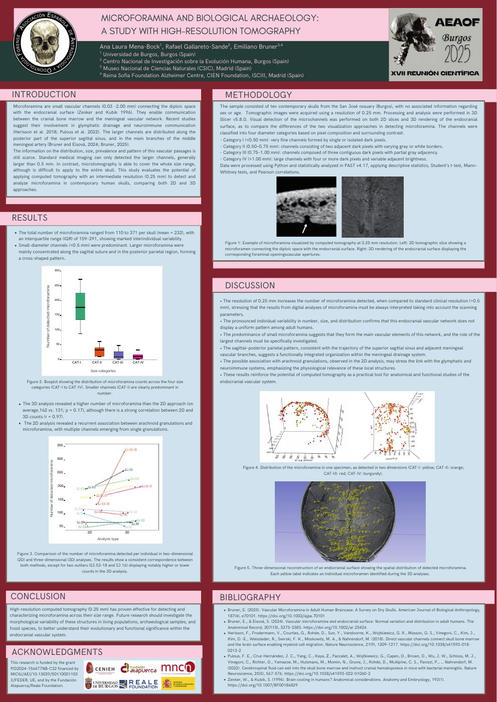
Microforamina and biological archaeology: a study with high-resolution tomographyMena-Bock, A. L., Gallareto-Sande, R., Bruner, E.
Resumen

Introduction. Microforamina are small vascular channels distributed on the endocranial surface of the human vault, with a size ranging from 0.02 to 2.00 mm. The larger ones are distributed in correspondence with the superior sagittal sinus and the larger branches of the middle meningeal vessels. They are possibly involved in cerebral immune responses and inflammation, as well as endocranial thermal regulation.

Objectives. The anatomy, distribution, and functions of these channels are still poorly known. Medical tomographic scans only provide information on the distribution of the larger channels. Microtomography involves technical and sample limitations. In this survey, we investigate the distribution and morphology of these microforamina on a cranial collection from an archaeological sample with a resolution that is intermediate between medical and microtomographic machines.

Material and methods. Ten individuals from a Spanish archaeological collection were scanned at a resolution of 0.25 mm. Microforamina have been detected on 2D sections, as well as on 3D reconstructions. Results are described in terms of anatomical distribution and channel size.

Results and discussion. The current resolution is able to detect a large number of channels, namely 100-350 per individual. Most individuals present between 150 and 250 channels. The largest microforamina can exceed 2 mm in lumen size. These results suggest that CT scanning cranial specimens with a resolution of 0.25 mm is an acceptable trade-off for studies in bioarchaeology and forensic anthropology.

XVII Reunión Científica de la AEAOF
Burgos, España
2024Póster
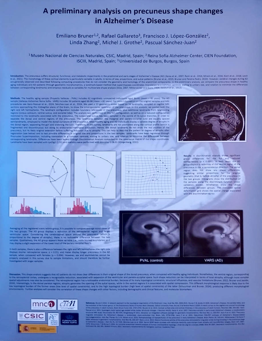
A preliminary analysis on precuneus shape changes in Alzheimer's DiseaseBruner, E., Gallareto-Sande, R., López-González, F. J., Zhang, L., Grothe, M. J., Sánchez-Juan, P.
Resumen

The precuneus suffers structural, functional, and metabolic impairments during the prodromal and early stages of Alzheimer's Disease (AD). However, most of the information on the corresponding anatomical changes comes from imaging methods that do not preserve the original morphology and homology of the involved cerebral regions. In this preliminary study, we consider whether some of these changes can be recognized in terms of gross geometry and spatial arrangement. We compare a sample of non-AD aging individuals with a sample of patients diagnosed with Alzheimer’s Disease. Geometric morphometrics was used to analyse shape differences between groups, through a landmark-based model including the precuneus boundaries and other neighboring references. After registration, differences between the two groups in the dorsal region, in terms of gross atrophy, are not patent. Instead, in AD patients there is a significant reduction of the ventral region, namely those parts contiguous with the retrosplenial cortex. These results are interpreted in terms of brain topology and morphological burden of these areas.

International Congress on Neurodegenerative Diseases
Lisboa, Portugal